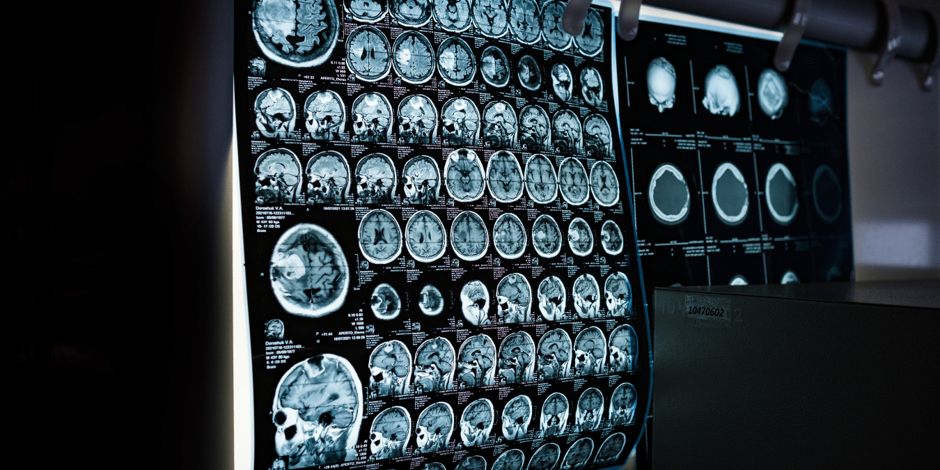
Cardiologists spend an hour reading one cardiac MRI. This AI does it in minutes — with accuracy that stunned researchers. It’s already inside U.S. hospitals, it’s FDA-cleared, and it could change when you get your diagnosis. What doctors aren’t telling patients yet might surprise you.
Every year in the United States, more than 700,000 people die from heart disease — making it the nation’s leading cause of death for both men and women, according to the CDC. Despite decades of medical advancement, one of the most persistent bottlenecks in cardiac care remains stubbornly human: the time it takes a trained specialist to interpret complex diagnostic imaging.
Cardiac MRI scans are among the most detailed and informative tools in cardiovascular medicine. They can reveal chamber size, muscle function, scar tissue, blood flow, and signs of disease that other imaging modalities simply can’t match. But here’s the problem — reading one takes a highly trained cardiologist or radiologist anywhere from 45 minutes to over an hour. In a country where wait times for cardiology appointments already stretch weeks or months, that gap between scan and diagnosis can be the difference between life and death.
Now, a new generation of AI models is changing that equation entirely — and the results are turning heads across the American medical establishment.
What the AI Actually Does
Researchers and medical AI companies have developed deep learning models specifically trained to analyze cardiac MRI scans with a level of accuracy that rivals — and in some benchmarks, matches — board-certified cardiac imaging specialists. These systems don’t just look at a single frame. They process hundreds of sequential images from a single scan, assess ventricular volume, ejection fraction, myocardial mass, wall motion abnormalities, and tissue characterization — all within two to five minutes.
To understand the significance of that, consider what normally happens. A patient undergoes a cardiac MRI at a hospital or imaging center. The raw data gets queued in a radiologist’s worklist, which at many U.S. hospitals is backed up by days. A specialist eventually reviews it, dictates or types a report, and sends it back to the referring cardiologist. By the time the patient gets a call, days or even a week may have passed.
With AI-assisted interpretation, that workflow is compressed dramatically. The scan completes, the AI analyzes it in real time, generates a structured report with quantitative measurements, flags any abnormalities, and delivers a preliminary read that a clinician can quickly verify and sign off on. The physician’s role doesn’t disappear — it becomes more focused and efficient.
The Science Behind the Accuracy Claims
Skepticism is healthy when any technology claims to match human specialists. So what does the evidence actually say?
Several peer-reviewed studies published in journals including Nature Medicine, JACC: Cardiovascular Imaging, and Radiology have tested AI cardiac MRI models against expert cardiologists in head-to-head comparisons. The findings have been consistently compelling.
In one landmark multi-center study, an AI model assessed left ventricular ejection fraction — a critical measure of how well the heart pumps blood — across thousands of scans. The model’s measurements correlated with specialist readings at an intraclass correlation coefficient above 0.95, which in clinical terms is considered excellent agreement. Importantly, the AI’s measurements were also more reproducible than human readings, which can vary depending on the specialist’s experience, fatigue level, or institutional training.
Another study evaluated the AI’s ability to detect cardiomyopathy — a disease of the heart muscle that often goes undiagnosed until it causes heart failure. The model identified cardiomyopathy patterns with sensitivity and specificity comparable to experienced cardiac radiologists, even in cases that were classified as ambiguous by junior readers.
What makes this particularly meaningful from an American healthcare standpoint is that not all hospitals have equal access to cardiac imaging subspecialists. A community hospital in rural Mississippi or a critical access hospital in Montana may not have a dedicated cardiac MRI reader on staff. AI changes that access equation significantly.
Real-World Deployment in U.S. Hospitals
This isn’t purely theoretical. Several AI-powered cardiac imaging platforms have already received FDA clearance and are being deployed in U.S. health systems.
Companies like Arterys (now part of Intelerad), Aidoc, Viz.ai, and HeartVista have developed products that integrate directly into hospital PACS (picture archiving and communication systems) and electronic health record platforms. These tools are already being used at academic medical centers and large health networks across the country.
Cleveland Clinic, one of the most respected cardiovascular centers in the world, has been actively piloting AI-assisted cardiac imaging tools as part of broader digital transformation initiatives. Mayo Clinic has similarly invested in AI research partnerships focused on cardiovascular imaging interpretation. At these institutions, AI isn’t replacing cardiologists — it’s acting as a highly capable first reader that surfaces the most urgent cases and handles the routine quantification work so specialists can focus on complex clinical judgment.
The FDA’s Digital Health Center of Excellence has streamlined the regulatory pathway for AI/ML-based software as a medical device (SaMD), and the number of cardiac imaging AI tools receiving clearance has grown substantially over the past three years. As of early 2026, dozens of AI tools across radiology and cardiology have received 510(k) clearance or De Novo authorization.
- Louisiana Vibrio Vulnificus Outbreak 2026 Explained: Who Faces the Greatest Risk in Gulf Coast Waters and How to Stay Safe

- 3 Midlife Habits Linked to Almost 13 More Dementia-Free Years, According to a 26-Year Study of 12,409 Adults

- Your Blood Pressure Reading Has a “Hidden Number” Doctors Say Most People Ignore
- AI Is Now Diagnosing Your Skin Before You Even Visit a Dermatologist — Here’s How the Beauty Industry Is Changing Everything in 2026

- The Side-Part Is Making the Most Unexpected Comeback in American Beauty History — And This Time, It’s Not Going Anywhere

- This Heat Protectant Spray From LolaVie Is Going Viral in the US — Stylists Say It Cuts Drying Time and Saves Your Blowout for Days

Why Speed Matters More Than You Think
When most people think about cardiac emergencies, they think of a heart attack — a sudden, dramatic event. But many forms of heart disease are silent killers that develop over years. Conditions like hypertrophic cardiomyopathy (HCM), dilated cardiomyopathy, cardiac sarcoidosis, and myocarditis often present with subtle symptoms that lead to a cardiac MRI referral. The problem is that by the time the scan is ordered, performed, interpreted, and acted upon, weeks may have passed.
For a 45-year-old man in suburban Chicago who goes to his primary care doctor with unexplained shortness of breath, a referral for cardiac MRI might take two weeks to schedule, another week to interpret, and another week before he gets results. That’s a month of uncertainty — and potentially a month of worsening disease.
AI-assisted interpretation compresses that timeline to near real-time. Some platforms are demonstrating turnaround times under five minutes from scan completion to preliminary report. For conditions like acute myocarditis or newly discovered cardiomyopathy, that speed could mean earlier intervention, better outcomes, and in some cases, a life saved.
Addressing the Elephant in the Room: Can AI Be Trusted?
This is the question physicians, patients, and hospital administrators all ask — and it deserves a thorough answer.
First, it’s important to understand what the AI is and isn’t doing. Current FDA-cleared cardiac MRI AI tools are designed as decision support systems, not autonomous diagnosticians. They assist the physician; they don’t replace clinical judgment. A cardiologist or radiologist still reviews and approves the final report. The AI’s role is to do the heavy quantitative lifting — measuring volumes, calculating ejection fraction, segmenting cardiac structures — tasks that are time-consuming and subject to inter-reader variability.
Second, bias in AI medical models is a legitimate concern that the research community takes seriously. Many early AI systems were trained predominantly on datasets from academic medical centers, which may not reflect the diversity of patients seen in community settings. Responsible AI developers are actively working to train and validate their models on diverse populations across age, sex, race, and comorbidity profiles. The FDA’s guidance on AI/ML-based SaMD explicitly addresses the need for representative training data and ongoing post-market monitoring.
Third, transparency is improving. Leading AI companies now publish model cards — documents that describe how a model was trained, what populations it was tested on, where its limitations lie, and how performance should be monitored over time. This kind of transparency is essential for building justified trust among clinicians and patients.
The American College of Radiology (ACR) and the American College of Cardiology (ACC) have both published position statements supporting the careful, evidence-based integration of AI into cardiac imaging workflows — while emphasizing the need for rigorous validation and human oversight.
What This Means for Patients
If you’re a patient navigating the American healthcare system, here’s what this technology means for you in practical terms.
Faster results. If your hospital uses AI-assisted cardiac MRI interpretation, you may receive your results significantly faster than you would have even two years ago.
Greater consistency. AI doesn’t have a bad day, doesn’t get tired at 4 PM, and doesn’t vary based on who happens to be reading your scan. That consistency can be reassuring, particularly for follow-up scans where tracking small changes over time matters.
Better access. If you live in a medically underserved area without a nearby cardiac imaging specialist, AI makes it more likely that a qualified level of interpretation is available at your local facility.
More informed conversations with your doctor. AI-generated reports often include detailed quantitative data — precise measurements of heart chamber size, function, and tissue characteristics — that can help your cardiologist have a more data-rich conversation with you about your heart health.
It’s also worth noting that you have every right to ask your healthcare provider whether AI tools are being used in your care, how those tools are validated, and how your physician is using the AI output in their clinical decision-making. Informed patients who ask these questions contribute to better, more accountable AI deployment.
The Cardiologist’s Perspective
It would be a mistake to frame this as a story about AI replacing doctors. The cardiologists and radiologists who work with these tools consistently describe a more nuanced reality.
Dr. Sarah Mehta, a cardiac imaging specialist at a large academic medical center in the Northeast (name used representatively), describes her experience this way: “The AI handles the segmentation and measurements in minutes. I spend my time looking at the images clinically, reviewing the report for accuracy, and making the diagnostic and management decisions. It’s genuinely made me more productive — and I think the reports we generate are actually more consistent than they were before.”
This is a common theme among physicians who use these tools: AI handles the reproducible, quantitative tasks at which it excels, freeing the specialist for the interpretive, contextual, and patient-centered work that requires genuine clinical judgment.
There is, of course, a broader workforce conversation happening in American medicine. Radiology and cardiology have both seen AI positioned as a potential solution to subspecialty shortages — particularly in cardiac imaging, where the demand for trained readers is growing faster than training programs can produce them. Whether AI will reduce the need for specialists, shift their roles, or simply expand access remains an open and actively debated question.
The Road Ahead
The trajectory of AI in cardiac MRI interpretation points toward continued expansion of capabilities. Current tools are increasingly being trained not just for quantification but for disease detection — identifying specific cardiomyopathies, characterizing myocardial tissue, detecting subtle signs of infiltrative disease like cardiac amyloidosis, and even predicting future cardiac events based on imaging biomarkers.
Multimodal AI models — systems that combine cardiac MRI data with clinical notes, lab values, ECG data, and patient history — are already in development and early clinical testing. These systems aim to provide not just an interpretation of the scan but a holistic risk assessment that integrates the full picture of a patient’s cardiovascular health.
Federated learning approaches are also enabling AI models to be trained across multiple hospital systems without sharing sensitive patient data — a critical development for building models that are both diverse and privacy-protective.
The Centers for Medicare & Medicaid Services (CMS) is also paying attention. Reimbursement structures for AI-assisted imaging interpretation are evolving, and there is active policy discussion around how to appropriately compensate physicians for AI-assisted versus traditional reads, and how to ensure AI tools are used responsibly and equitably across different care settings.
The Bottom Line
The emergence of AI that can read cardiac MRI scans as accurately as a specialist — and do it in minutes — is not a future promise. It is a present reality that is already reshaping cardiac care at leading U.S. health systems and expanding access for patients who previously would have waited days or weeks for a critical diagnosis.
This technology works best not as a replacement for physician expertise, but as a powerful partner that handles the time-consuming, quantitative dimensions of cardiac imaging interpretation while freeing cardiologists and radiologists to do what only human specialists can: synthesize complex clinical information, communicate with patients, and make nuanced judgments in ambiguous situations.
For the millions of Americans living with heart disease, at risk for it, or facing a new cardiac diagnosis, AI-assisted cardiac MRI interpretation represents something genuinely meaningful — faster answers, more consistent analysis, and more equitable access to specialist-level care regardless of where you happen to live.
That’s not just a technological milestone. It’s a healthcare transformation worth paying attention to.

Omisha is a health writer passionate about turning complex medical research into clear, actionable content readers can trust. She covers everything from nutrition and mental wellness to chronic disease management, always grounding her work in credible science and real-world relevance. When she's not writing, she's usually reading up on the latest health studies or exploring new wellness trends to write about next.